
Anti-KCNMA1 Antibody

Catalog# ER1902-04
Anti-KCNMA1 Antibody
-
WB
-
IF-Cell
-
FC
-
Human
-
Mouse
-
Rabbit
-
unconjugated
Overview
Product Name
Anti-KCNMA1 Antibody
Antibody Type
Rabbit Polyclonal Antibody
Immunogen
Synthetic peptide within rat KCNMA1 aa 200-300.
Species Reactivity
Human, Mouse, Rabbit
Validated Applications
WB, IF-Cell, FC
Target Molecular Weight
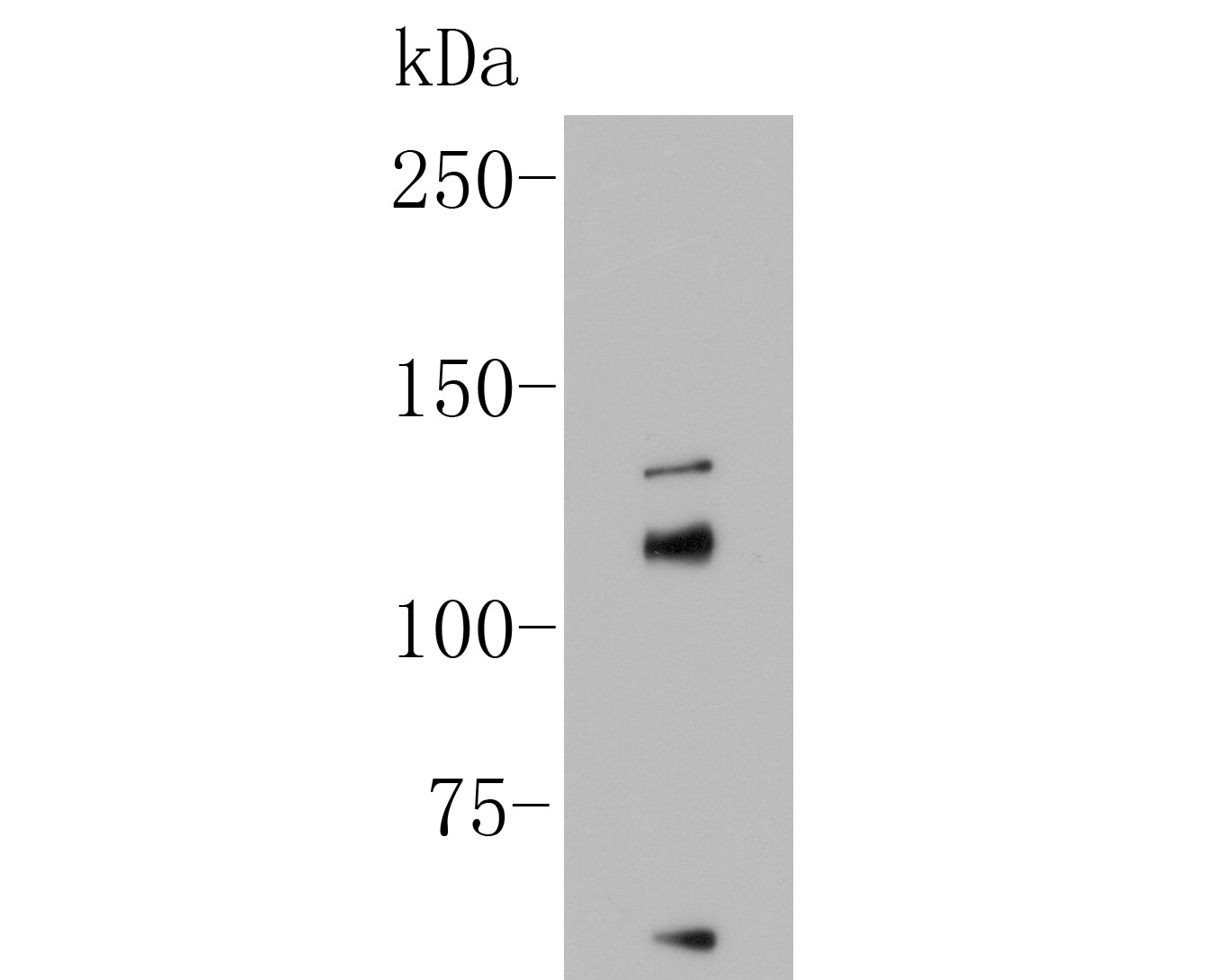
Predicted band size 138 kDa
Positive Control
A549 cell lysates, A549, PC-3M, HCT116.
Conjugation
unconjugated
RRID
Product Features
Form
Liquid
Concentration
1 mg/ml.(The concentration of this product may be batch-dependent)
Storage Instructions
Store at +4℃ after thawing. Aliquot store at -20℃. Avoid repeated freeze / thaw cycles.
Storage Buffer
1*PBS (pH7.4), 0.2% BSA, 50% Glycerol. Preservative: 0.05% Sodium Azide.
Isotype
IgG
Purification Method
Immunogen affinity purified.
Application Dilution
-
WB
-
1:500
-
IF-Cell
-
1:50-1:200
-
FC
-
1:50-1:100
Target
Function
Potassium channel activated by both membrane depolarization or increase in cytosolic Ca2+ that mediates export of K+. It is also activated by the concentration of cytosolic Mg2+. Its activation dampens the excitatory events that elevate the cytosolic Ca2+ concentration and/or depolarize the cell membrane. It therefore contributes to repolarization of the membrane potential. Plays a key role in controlling excitability in a number of systems, such as regulation of the contraction of smooth muscle, the tuning of hair cells in the cochlea, regulation of transmitter release, and innate immunity. In smooth muscles, its activation by high level of Ca2+, caused by ryanodine receptors in the sarcoplasmic reticulum, regulates the membrane potential. In cochlea cells, its number and kinetic properties partly determine the characteristic frequency of each hair cell and thereby helps to establish a tonotopic map. Kinetics of KCNMA1 channels are determined by alternative splicing, phosphorylation status and its combination with modulating beta subunits. Highly sensitive to both iberiotoxin (IbTx) and charybdotoxin (CTX).
Background References
1. Liang L. et. al. De novo loss-of-function KCNMA1 variants are associated with a new multiple malformation syndrome and a broad spectrum of developmental and neurological phenotypes. Hum Mol Genet. 2019 Jun 1.
2. Ibrahim S. et. al. Expression Profiling of Calcium Channels and Calcium-Activated Potassium Channels in Colorectal Cancer. Cancers (Basel). 2019 Apr 19;11(4).
Sequence Similarity
Belongs to the potassium channel family. Calcium-activated (TC 1.A.1.3) subfamily. KCa1.1/KCNMA1 sub-subfamily.
Tissue Specificity
Widely expressed. Except in myocytes, it is almost ubiquitously expressed.
Post-translational Modification
Phosphorylated (Probable). Phosphorylation by kinases such as PKA and/or PKG. In smooth muscles, phosphorylation affects its activity.; Palmitoylation by ZDHHC22 and ZDHHC23 within the intracellular linker between the S0 and S1 transmembrane domains regulates localization to the plasma membrane. Depalmitoylated by LYPLA1 and LYPLAL1, leading to retard exit from the trans-Golgi network.
Subcellular Location
Cell membrane.
UNIPROT
Synonyms
subfamily M subunit alpha-1 antibody
BK channel antibody
BKCA alpha antibody
BKCA alpha subunit antibody
BKTM antibody
Calcium-activated potassium channel antibody
Calcium-activated potassium channel subunit alpha-1 antibody
Drosophila slowpoke like antibody
hSlo antibody
K(VCA)alpha antibody
ExpandImages
-
Western blot analysis of on A549 cell lysate. Proteins were transferred to a PVDF membrane and blocked with 5% BSA in PBS for 1 hour at room temperature. The primary antibody (ER1902-04, 1/500) was used in 5% BSA at room temperature for 2 hours. Goat Anti-Rabbit IgG - HRP Secondary Antibody (HA1001) at 1:5,000 dilution was used for 1 hour at room temperature.
-
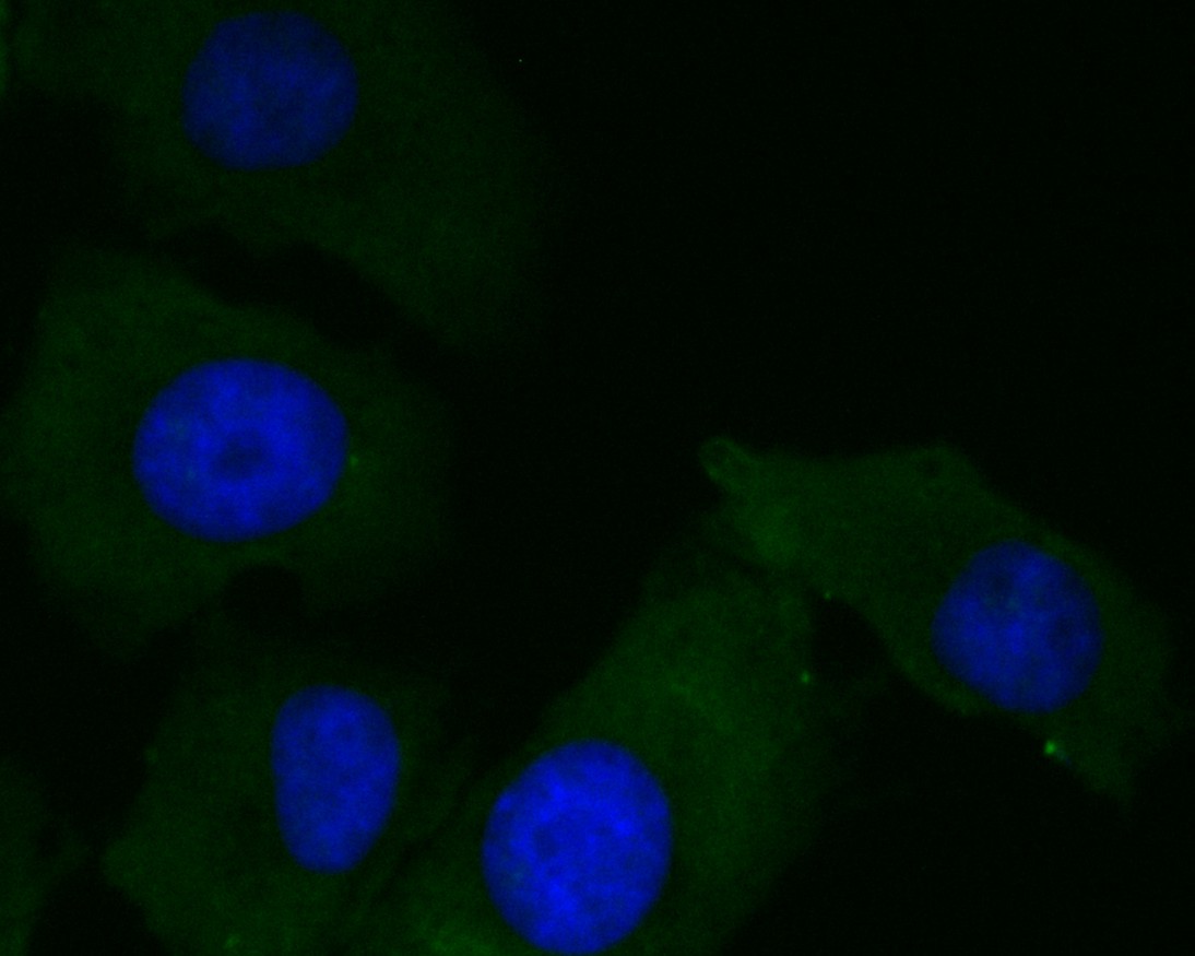
ICC staining of KCNMA1 in A549 cells (green). Formalin fixed cells were permeabilized with 0.1% Triton X-100 in TBS for 10 minutes at room temperature and blocked with 1% Blocker BSA for 15 minutes at room temperature. Cells were probed with the primary antibody (ER1902-04, 1/100) for 1 hour at room temperature, washed with PBS. Alexa Fluor®488 Goat anti-Rabbit IgG was used as the secondary antibody at 1/100 dilution. The nuclear counter stain is DAPI (blue)
-
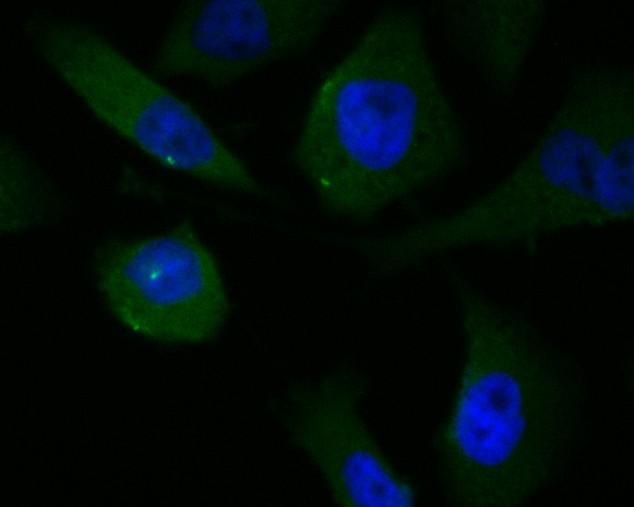
ICC staining of KCNMA1 in PC-3M cells (green). Formalin fixed cells were permeabilized with 0.1% Triton X-100 in TBS for 10 minutes at room temperature and blocked with 1% Blocker BSA for 15 minutes at room temperature. Cells were probed with the primary antibody (ER1902-04, 1/100) for 1 hour at room temperature, washed with PBS. Alexa Fluor®488 Goat anti-Rabbit IgG was used as the secondary antibody at 1/100 dilution. The nuclear counter stain is DAPI (blue)
-
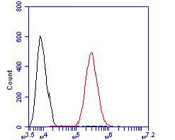
Flow cytometric analysis of KCNMA1 was done on HCT116 cells. The cells were fixed, permeabilized and stained with the primary antibody (ER1902-04, 1/100) (red). After incubation of the primary antibody at room temperature for an hour, the cells were stained with a Alexa Fluor 488-conjugated goat anti-rabbit IgG Secondary antibody at 1/500 dilution for 30 minutes.Unlabelled sample was used as a control (cells without incubation with primary antibody; black).
Please note: All products are "FOR RESEARCH USE ONLY AND ARE NOT INTENDED FOR DIAGNOSTIC OR THERAPEUTIC USE"