Collagen VI Recombinant Rabbit Monoclonal Antibody [SD83-03]

Overview
Product Name
Collagen VI Recombinant Rabbit Monoclonal Antibody [SD83-03]
Antibody Type
Recombinant Rabbit monoclonal Antibody
Immunogen
Recombinant protein within Human Collagen VI alpha1 aa 17-255 / 1,028.
Species Reactivity
Human, Mouse, Rat
Validated Applications
WB, IF-Cell, IF-Tissue, IHC-P
Target Molecular Weight
Predicted band size: 109 kDa
Positive Control
NIH/3T3 cell lysate, Human kidney tissue lysate, Rat heart tissue lysate, Rat kidney tissue lysate, Saos-2 cell lysate, C6 cell lysate, NIH/3T3, human liver tissue, human colon tissue, human skin tissue, mouse colon tissue, rat heart tissue, rat kidney tissue.
Conjugation
unconjugated
Clone Number
SD83-03
RRID
Product Features
Form
Liquid
Concentration
1 mg/mL.(The concentration of this product may be batch-dependent)
Storage Instructions
Shipped at 4℃. Store at +4℃ short term (1-2 weeks). Store at -20℃ long term.
Storage Buffer
1*PBS (pH7.4), 0.1% BSA, 40% Glycerol, 0.2% Proclean 950.
Isotype
IgG
Purification Method
Protein A affinity purified.
Application Dilution
-
WB
-
1:2,000
-
IF-Cell
-
1:100-1:500
-
IF-Tissue
-
1:100-1:500
-
IHC-P
-
1:400-1:1,000
Target
Function
Collagen VI (ColVI) is a type of collagen primarily associated with the extracellular matrix of skeletal muscle. ColVI maintains regularity in muscle function and stabilizes the cell membrane. It is synthesized by a complex, multistep pathway that leads to the formation of a unique network of linked microfilaments located in the extracellular matrix (ECM). ColVI plays a vital role in numerous cell types, including chondrocytes, neurons, myocytes, fibroblasts, and cardiomyocytes. ColVI molecules are made up of three alpha chains: α1(VI), α2(VI), and α3(VI). It is encoded by 6 genes: COL6A1, COL6A2, COL6A3, COL6A4, COL6A5, and COL6A6. The chain lengths of α1(VI) and α2(VI) are about 1,000 amino acids. The chain length of α3(VI) is roughly a third larger than those of α1(VI) and α2(VI), and it consists of several spliced variants within the range of 2,500 to 3,100 amino acids. The first two alpha chains subunits of ColVI have a molecular weight of 140-150 KDa and the third polypeptide chain is larger with a molecular weight of 250-300kDa. ColVI is also found in the skin, lungs, blood vessels, cornea and intervertebral disc. It also forms part of the peripheral nerves, brain, myocardium and adipose tissue.
Background References
1. Kristofik N et al. Impaired von Willebrand factor adhesion and platelet response in thrombospondin-2 knockout mice. Blood 128:1642-50 (2016).
2. Okawa S et al. Lipopolysaccharide induces expression of collagen VI in the rat lung. J Toxicol Pathol 28:37-41 (2015).
Sequence Similarity
Belongs to the type VI collagen family.
Post-translational Modification
Prolines at the third position of the tripeptide repeating unit (G-X-Y) are hydroxylated in some or all of the chains.
Subcellular Location
Extracellular matrix.
UNIPROT
Synonyms
Alpha 1 (VI) chain (61 AA) antibody
CO6A1_HUMAN antibody
COL6A1 antibody
Collagen alpha-1(VI) chain antibody
Collagen type VI alpha 1 antibody
Collagen VI alpha 1 polypeptide antibody
CollagenVI antibody
Human mRNA for collagen VI alpha 2 C terminal globular domain antibody
OPLL antibody
PP3610 antibody
Images
-

Western blot analysis of Collagen VI on different lysates with Rabbit anti-Collagen VI antibody (ET1612-91) at 1/2,000 dilution.
Lane 1: NIH/3T3 cell lysate (15 µg/Lane)
Lane 2: Human kidney tissue lysate (20 µg/Lane)
Lane 3: Rat heart tissue lysate (20 µg/Lane)
Lane 4: Rat kidney tissue lysate (20 µg/Lane)
Predicted band size: 109 kDa
Observed band size: 145 kDa
Exposure time: 1 minute 30 seconds;
4-20% SDS-PAGE gel.
Proteins were transferred to a PVDF membrane and blocked with 5% NFDM/TBST for 1 hour at room temperature. The primary antibody (ET1612-91) at 1/2,000 dilution was used in 5% NFDM/TBST at 4℃ overnight. Goat Anti-Rabbit IgG - HRP Secondary Antibody (HA1001) at 1/50,000 dilution was used for 1 hour at room temperature. -

Western blot analysis of Collagen VI on different lysates with Rabbit anti-Collagen VI antibody (ET1612-91) at 1/1,000 dilution.
Lane 1: Saos-2 cell lysate
Lane 2: NIH/3T3 cell lysate
Lane 3: C6 cell lysate
Lysates/proteins at 20 µg/Lane.
Predicted band size: 145 kDa
Observed band size: 145 kDa
Exposure time: 6 seconds;
4-20% SDS-PAGE gel.
Proteins were transferred to a PVDF membrane and blocked with 5% NFDM/TBST for 1 hour at room temperature. The primary antibody (ET1612-91) at 1/1,000 dilution was used in 5% NFDM/TBST at 4℃ overnight. Goat Anti-Rabbit IgG - HRP Secondary Antibody (HA1001) at 1/50,000 dilution was used for 1 hour at room temperature. -

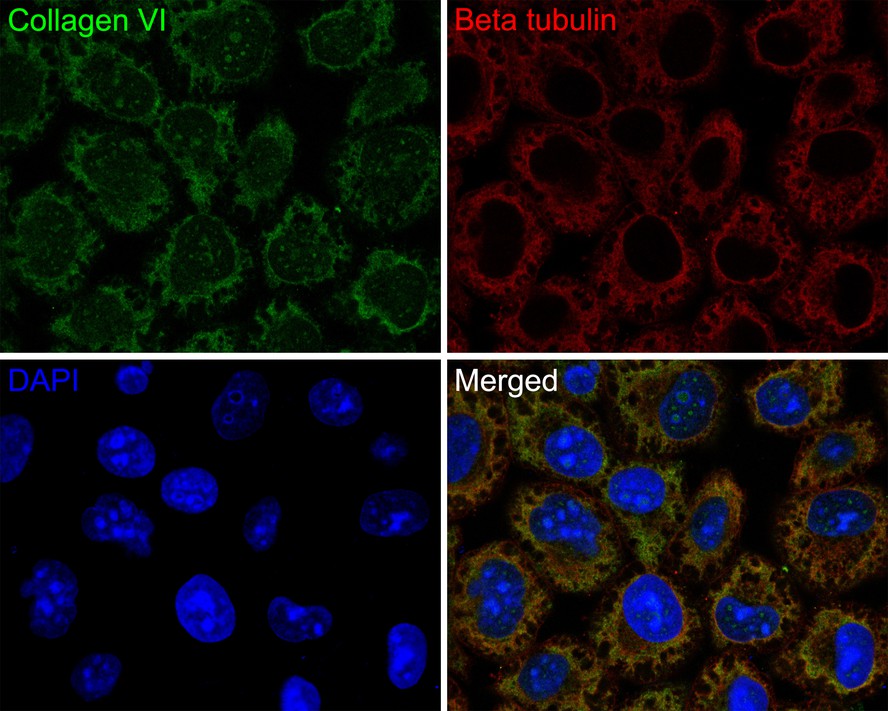
Immunocytochemistry analysis of NIH/3T3 cells labeling Collagen VI with Rabbit anti-Collagen VI antibody (ET1612-91) at 1/100 dilution.
Cells were fixed in 4% paraformaldehyde for 20 minutes at room temperature, permeabilized with 0.1% Triton X-100 in PBS for 5 minutes at room temperature, then blocked with 1% BSA in 10% negative goat serum for 1 hour at room temperature. Cells were then incubated with Rabbit anti-Collagen VI antibody (ET1612-91) at 1/100 dilution in 1% BSA in PBST overnight at 4 ℃. Goat Anti-Rabbit IgG H&L (iFluor™ 488, HA1121) was used as the secondary antibody at 1/1,000 dilution. PBS instead of the primary antibody was used as the secondary antibody only control. Nuclear DNA was labelled in blue with DAPI.
Beta tubulin (M1305-2, red) was stained at 1/100 dilution overnight at +4℃. Goat Anti-Mouse IgG H&L (iFluor™ 594, HA1126) was used as the secondary antibody at 1/1,000 dilution. -

Immunohistochemical analysis of paraffin-embedded human liver tissue with Rabbit anti-Collagen VI antibody (ET1612-91) at 1/1,000 dilution.
The section was pre-treated using heat mediated antigen retrieval with Tris-EDTA buffer (pH 9.0) for 20 minutes. The tissues were blocked in 1% BSA for 20 minutes at room temperature, washed with ddH2O and PBS, and then probed with the primary antibody (ET1612-91) at 1/1,000 dilution for 1 hour at room temperature. The detection was performed using an HRP conjugated compact polymer system. DAB was used as the chromogen. Tissues were counterstained with hematoxylin and mounted with DPX. -

Immunohistochemical analysis of paraffin-embedded human colon tissue with Rabbit anti-Collagen VI antibody (ET1612-91) at 1/400 dilution.
The section was pre-treated using heat mediated antigen retrieval with Tris-EDTA buffer (pH 9.0) for 20 minutes. The tissues were blocked in 1% BSA for 20 minutes at room temperature, washed with ddH2O and PBS, and then probed with the primary antibody (ET1612-91) at 1/400 dilution for 1 hour at room temperature. The detection was performed using an HRP conjugated compact polymer system. DAB was used as the chromogen. Tissues were counterstained with hematoxylin and mounted with DPX. -

Immunohistochemical analysis of paraffin-embedded human skin tissue with Rabbit anti-Collagen VI antibody (ET1612-91) at 1/400 dilution.
The section was pre-treated using heat mediated antigen retrieval with Tris-EDTA buffer (pH 9.0) for 20 minutes. The tissues were blocked in 1% BSA for 20 minutes at room temperature, washed with ddH2O and PBS, and then probed with the primary antibody (ET1612-91) at 1/400 dilution for 1 hour at room temperature. The detection was performed using an HRP conjugated compact polymer system. DAB was used as the chromogen. Tissues were counterstained with hematoxylin and mounted with DPX. -

Immunohistochemical analysis of paraffin-embedded mouse colon tissue with Rabbit anti-Collagen VI antibody (ET1612-91) at 1/1,000 dilution.
The section was pre-treated using heat mediated antigen retrieval with Tris-EDTA buffer (pH 9.0) for 20 minutes. The tissues were blocked in 1% BSA for 20 minutes at room temperature, washed with ddH2O and PBS, and then probed with the primary antibody (ET1612-91) at 1/1,000 dilution for 1 hour at room temperature. The detection was performed using an HRP conjugated compact polymer system. DAB was used as the chromogen. Tissues were counterstained with hematoxylin and mounted with DPX. -

Immunohistochemical analysis of paraffin-embedded rat heart tissue with Rabbit anti-Collagen VI antibody (ET1612-91) at 1/1,000 dilution.
The section was pre-treated using heat mediated antigen retrieval with Tris-EDTA buffer (pH 9.0) for 20 minutes. The tissues were blocked in 1% BSA for 20 minutes at room temperature, washed with ddH2O and PBS, and then probed with the primary antibody (ET1612-91) at 1/1,000 dilution for 1 hour at room temperature. The detection was performed using an HRP conjugated compact polymer system. DAB was used as the chromogen. Tissues were counterstained with hematoxylin and mounted with DPX. -

Immunohistochemical analysis of paraffin-embedded rat kidney tissue with Rabbit anti-Collagen VI antibody (ET1612-91) at 1/1,000 dilution.
The section was pre-treated using heat mediated antigen retrieval with Tris-EDTA buffer (pH 9.0) for 20 minutes. The tissues were blocked in 1% BSA for 20 minutes at room temperature, washed with ddH2O and PBS, and then probed with the primary antibody (ET1612-91) at 1/1,000 dilution for 1 hour at room temperature. The detection was performed using an HRP conjugated compact polymer system. DAB was used as the chromogen. Tissues were counterstained with hematoxylin and mounted with DPX. -

Immunofluorescence analysis of paraffin-embedded human liver tissue labeling Collagen VI with Rabbit anti-Collagen VI antibody (ET1612-91) at 1/100 dilution.
The section was pre-treated using heat mediated antigen retrieval with Tris-EDTA buffer (pH 9.0) for 20 minutes. The tissues were blocked in 10% negative goat serum for 1 hour at room temperature, washed with PBS, and then probed with the primary antibody (ET1612-91, green) at 1/100 dilution overnight at 4 ℃, washed with PBS.
Goat Anti-Rabbit IgG H&L (iFluor™ 488, HA1121) was used as the secondary antibody at 1/1,000 dilution. Nuclei were counterstained with DAPI (blue). -

Immunofluorescence analysis of paraffin-embedded mouse colon tissue labeling Collagen VI with Rabbit anti-Collagen VI antibody (ET1612-91) at 1/100 dilution.
The section was pre-treated using heat mediated antigen retrieval with Tris-EDTA buffer (pH 9.0) for 20 minutes. The tissues were blocked in 10% negative goat serum for 1 hour at room temperature, washed with PBS, and then probed with the primary antibody (ET1612-91, green) at 1/100 dilution overnight at 4 ℃, washed with PBS.
Goat Anti-Rabbit IgG H&L (iFluor™ 488, HA1121) was used as the secondary antibody at 1/1,000 dilution. Nuclei were counterstained with DAPI (blue).
Please note: All products are "FOR RESEARCH USE ONLY AND ARE NOT INTENDED FOR DIAGNOSTIC OR THERAPEUTIC USE"
Citation
-
Luteolin Mitigates Photoaging Caused by UVA-Induced Fibroblast Senescence by Modulating Oxidative Stress Pathways
Journal: International Journal Of Molecular Sciences
DOI: 10.3390/ijms26051809
IF: 4.9
Application: WB
Reactivity: Mouse
Publish date: 2025 Feb
-
TGF-β1-induced collagen promotes chicken ovarian follicle development via an intercellular cooperative pattern
Journal: Cell Biology International
DOI:
IF: 3.613
Application: WB
Reactivity: Chicken
Publish date: 2021 Jun
-
Transcriptome profiling analysis of underlying regulation of growing follicle development in the chicken
Journal: Poultry Science
DOI:
IF: 2.659
Application: IHC-P,WB
Reactivity: Chicken
Publish date: 2020 Jun
Products with the same target and pathway

Collagen VI Recombinant Rabbit Monoclonal Antibody [SD83-03] - BSA and Azide free
Application: WB,IF-Cell,IF-Tissue,IHC-P
Reactivity: Human,Mouse,Rat
Conjugate: unconjugated