
Glutathione Peroxidase 1 Mouse Monoclonal Antibody [C5-F6]


Catalog# EM40708
Glutathione Peroxidase 1 Mouse Monoclonal Antibody [C5-F6]
-
WB
-
IF-Cell
-
IHC-P
-
FC
-
Human
-
unconjugated
Overview
Product Name
Glutathione Peroxidase 1 Mouse Monoclonal Antibody [C5-F6]
Antibody Type
Mouse Monoclonal Antibody
Immunogen
Recombinant protein within Human Glutathione Peroxidase 1 aa 25-203 / 203.
Species Reactivity
Human
Validated Applications
WB, IF-Cell, IHC-P, FC
Target Molecular Weight
Predicted band size: 22 kDa
Positive Control
THP-1 cell lysate, HEK-293 cell lysate, HepG2 cell lysate, SH-SY5Y cell lysate, HT-29 cell lysate, Hela, HepG2, human kidney tissue.
Conjugation
unconjugated
Clone Number
C5-F6
RRID
Product Features
Form
Liquid
Concentration
2 mg/mL.(The concentration of this product may be batch-dependent)
Storage Instructions
Shipped at 4℃. Store at +4℃ short term (1-2 weeks). Store at -20℃ long term.
Storage Buffer
1*PBS (pH7.4), 0.2% BSA, 40% Glycerol. Preservative: 0.05% Sodium Azide.
Isotype
IgG2b
Purification Method
Protein A affinity purified.
Application Dilution
-
WB
-
1:2,000
-
IHC-P
-
1:200
-
IF-Cell
-
1:200
-
FC
-
1:100-1:200
Target
Function
GPX1 is ubiquitously expressed in many tissues, where it protects cells from oxidative stress. Within cells, it localizes to the cytoplasm and mitochondria. As a glutathione peroxidase, GPx1 functions in the detoxification of hydrogen peroxide, specifically by catalyzing the reduction of hydrogen peroxide to water. The glutathione peroxidase also catalyzes the reduction of other organic hydroperoxides, such as lipid peroxides, to the corresponding alcohols. GPx1 typically uses glutathione (GSH) as the reductant, but when glutathione synthetase (GSS) is, as in brain mitochondria, γ-glutamylcysteine can serve as the reductant instead. The protein encoded by this gene protects from CD95-induced apoptosis in cultured breast cancer cells and inhibits 5-lipoxygenase in blood cells, and its overexpression delays endothelial cell death and increases resistance to toxic challenges, especially oxidative stress. This protein is one of only a few proteins known in higher vertebrates to contain selenocysteine, which occurs at the active site of glutathione peroxidase and is coded by the nonsense (stop) codon TGA.
Background References
1. "Association between the GCG polymorphism of the selenium dependent GPX1 gene and the risk of young onset prostate cancer." Kote-Jarai Z., Durocher F., Edwards S.M., Hamoudi R., Jackson R.A., Ardern-Jones A., Murkin A., Dearnaley D.P., Kirby R., Houlston R., Easton D.F., Eeles R. Prostate Cancer Prostatic Dis. 5:189-192(2002)
2. "Functional variants in the glutathione peroxidase-1 (GPX-1) gene are associated with increased intima-media thickness of carotid arteries and risk of macrovascular diseases in Japanese type 2 diabetic patients." Hamanishi T., Furuta H., Kato H., Doi A., Tamai M., Shimomura H., Sakagashira S., Nishi M., Sasaki H., Sanke T., Nanjo K. Diabetes 53:2455-2460(2004)
Sequence Similarity
Belongs to the glutathione peroxidase family.
Tissue Specificity
Expressed in platelets (at protein level).
Post-translational Modification
During periods of oxidative stress, Sec-49 may react with a superoxide radical, irreversibly lose hydroselenide and be converted to dehydroalanine.
Subcellular Location
Cytoplasm.
UNIPROT
Synonyms
AL033363 antibody
Cellular glutathione peroxidase antibody
Glutathione peroxidase 1 antibody
Glutathione peroxidase antibody
GPx 1 antibody
GPx-1 antibody
GPX1 antibody
GPX1_HUMAN antibody
GPXD antibody
GSHPx-1 antibody
ExpandImages
-
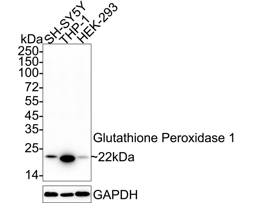
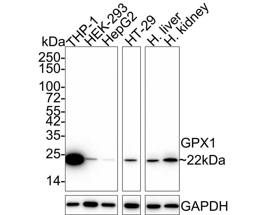
Western blot analysis of Glutathione Peroxidase 1 on different lysates with Mouse anti-Glutathione Peroxidase 1 antibody (EM40708) at 1/2,000 dilution.
Lane 1: THP-1 cell lysate
Lane 2: HEK-293 cell lysate
Lane 3: HepG2 cell lysate
Lane 4: SH-SY5Y cell lysate
Lane 5: HT-29 cell lysate
Lysates/proteins at 20 µg/Lane.
Predicted band size: 22 kDa
Observed band size: 22 kDa
Exposure time: 10 seconds; ECL: K1801;
4-20% SDS-PAGE gel.
Proteins were transferred to a PVDF membrane and blocked with 5% NFDM/TBST for 1 hour at room temperature. The primary antibody (EM40708) at 1/2,000 dilution was used in 5% NFDM/TBST at 4℃ overnight. Goat Anti-Mouse IgG - HRP Secondary Antibody (HA1006) at 1/50,000 dilution was used for 1 hour at room temperature. -
ICC staining GPX1 in Hela cells (red). Cells were fixed in paraformaldehyde, permeabilised with 0.25% Triton X100/PBS.
-
ICC staining GPX1 in HepG2 cells (red). Cells were fixed in paraformaldehyde, permeabilised with 0.25% Triton X100/PBS.
-
Immunohistochemical analysis of paraffin-embedded human kidney tissue using anti-GPX1 antibody. Counter stained with hematoxylin.
Please note: All products are "FOR RESEARCH USE ONLY AND ARE NOT INTENDED FOR DIAGNOSTIC OR THERAPEUTIC USE"
Alternative Products

Glutathione peroxidase 1 Recombinant Mouse Monoclonal Antibody [C5-A10-R]
Application: WB,IF-Cell,IHC-P,FC
Reactivity: Human
Conjugate: unconjugated
Glutathione Peroxidase 1 Recombinant Rabbit Monoclonal Antibody [JF0944]
Application: WB,IF-Tissue,IHC-P
Reactivity: Human,Mouse
Conjugate: unconjugated
Glutathione peroxidase 1 Recombinant Mouse Monoclonal Antibody [A6-C0-B9-R]
Application: WB,IF-Cell,IHC-P,FC
Reactivity: Human
Conjugate: unconjugated

Glutathione Peroxidase 1 Recombinant Rabbit Monoclonal Antibody [JJ092-07]
Application: WB,IP
Reactivity: Human,Mouse,Rat
Conjugate: unconjugated
Products with the same target and pathway
Glutathione peroxidase 1 Recombinant Mouse Monoclonal Antibody [C5-A10-R]
Application: WB,IF-Cell,IHC-P,FC
Reactivity: Human
Conjugate: unconjugated
Glutathione Peroxidase 1 Recombinant Rabbit Monoclonal Antibody [JF0944]
Application: WB,IF-Tissue,IHC-P
Reactivity: Human,Mouse
Conjugate: unconjugated

Glutathione peroxidase 1 Mouse Monoclonal Antibody [C5-A10]
Application: WB,IF-Cell,FC,IHC-P
Reactivity: Human,Mouse
Conjugate: unconjugated

Glutathione Peroxidase 1 Rabbit Polyclonal Antibody
Application: WB,IF-Cell,IHC-P,FC
Reactivity: Human,Mouse
Conjugate: unconjugated

Glutathione peroxidase 1 Recombinant Mouse Monoclonal Antibody [A6-C0-B9-R] - BSA and Azide free
Application: WB,IF-Cell,IHC-P,FC
Reactivity: Human
Conjugate: unconjugated

Glutathione peroxidase 1 Mouse Monoclonal Antibody [A6-C0-B9]
Application: WB,IHC-P,FC,IF-Cell
Reactivity: Human
Conjugate: unconjugated

Glutathione peroxidase 1 Recombinant Mouse Monoclonal Antibody [C5-A10-R] - BSA and Azide free
Application: WB,IF-Cell,IHC-P,FC
Reactivity: Human
Conjugate: unconjugated
Glutathione peroxidase 1 Recombinant Mouse Monoclonal Antibody [A6-C0-B9-R]
Application: WB,IF-Cell,IHC-P,FC
Reactivity: Human
Conjugate: unconjugated